

MedIA (Medical Image Analysis, IF: 11.148)为医学影像分析领域的顶级期刊。
1 S. Kevin. Zhou, H. N. Le, K. Luu, H. V. Nguyen, and N. Ayache, “Deep reinforcement learning in medical imaging: A literature review,” Medical Image Analysis, 2021: 102193.
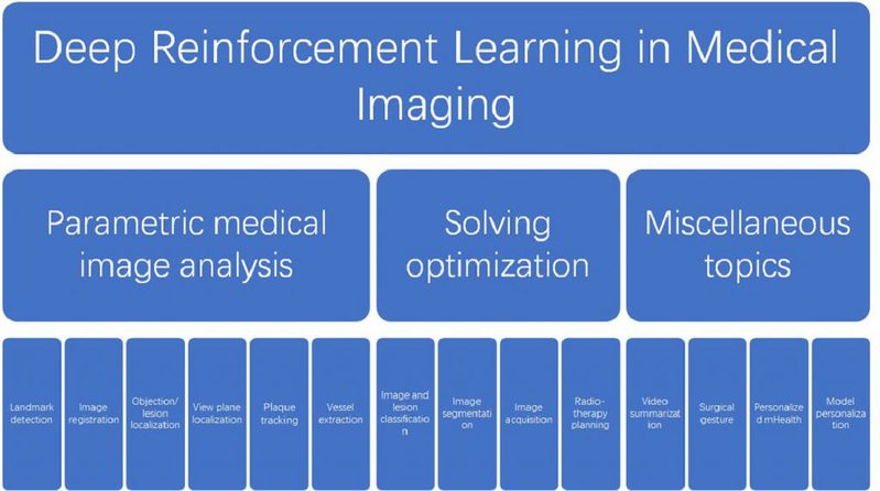
Abstract: Deep reinforcement learning (DRL) augments the reinforcement learning framework, which learns a sequence of actions that maximizes the expected reward, with the representative power of deep neural networks. Recent works have demonstrated the great potential of DRL in medicine and healthcare. This paper presents a literature review of DRL in medical imaging. We start with a comprehensive tutorial of DRL, including the latest model-free and model-based algorithms. We then cover existing DRL applications for medical imaging, which are roughly divided into three main categories: (i) parametric medical image analysis tasks including landmark detection, object/lesion detection, registration, and view plane localization; (ii) solving optimization tasks including hyperparameter tuning, selecting augmentation strategies, and neural architecture search; and (iii) miscellaneous applications including surgical gesture segmentation, personalized mobile health intervention, and computational model personalization. The paper concludes with discussions of future perspectives.
2 B. Zhou, X. Chen, S. Kevin Zhou, et al., “DuDoDR-Net: Dual-Domain Data Consistent Recurrent Network for Simultaneous Sparse View and Metal Artifact Reduction in Computed Tomography”, Medical Image Analysis, 2021: 102289.
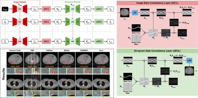
Abstract: Sparse-view computed tomography (SVCT) aims to reconstruct a cross-sectional image using a reduced number of x-ray projections. While SVCT can efficiently reduce the radiation dose, the reconstruction suffers from severe streak artifacts, and the artifacts are further amplified with the presence of metallic implants, which could adversely impact the medical diagnosis and other downstream applications. Previous methods have extensively explored either SVCT reconstruction without metallic implants, or full-view CT metal artifact reduction (MAR). The issue of simultaneous sparse-view and metal artifact reduction (SVMAR) remains under-explored, and it is infeasible to directly apply previous SVCT and MAR methods to SVMAR which may yield non-ideal reconstruction quality. In this work, we propose a dual-domain data consistent recurrent network, called DuDoDR-Net, for SVMAR. Our DuDoDR-Net aims to reconstruct an artifact-free image by recurrent image domain and sinogram domain restorations. To ensure the metal-free part of acquired projection data is preserved, we also develop the image data consistent layer (iDCL) and sinogram data consistent layer (sDCL) that are interleaved in our recurrent framework. Our experimental results demonstrate that our DuDoDR-Net is able to produce superior artifact-reduced results while preserving the anatomical structures, that outperform previous SVCT and SVMAR methods, under different sparse-view acquisition settings.
3 L. Han, Y. Lyu, C. Peng, and S. Kevin. Zhou, “Gan-based disentangle- ment learning for chest x-ray rib suppression,” Medical Image Analysis, 2022: 102369.
Abstract: Clinical evidence has shown that rib-suppressed chest X-rays (CXRs) can improve the reliability of pulmonary disease diagnosis. However, previous approaches on generating rib-suppressed CXR face challenges in preserving details and eliminating rib residues. We hereby propose a GAN-based disentanglement learning framework called Rib Suppression GAN, or RSGAN, to perform rib suppression by utilizing the anatomical knowledge embedded in unpaired computed tomography (CT) images. In this approach, we employ a residual map to characterize the intensity difference between CXR and the corresponding rib-suppressed result. To predict the residual map in CXR domain, we disentangle the image into structure- and contrast-specific features and transfer the rib structural priors from digitally reconstructed radiographs (DRRs) computed by CT. Furthermore, we employ additional adaptive loss to suppress rib residue and preserve more details. We conduct extensive experiments based on 1673 CT volumes, and four benchmarking CXR datasets, totaling over 120K images, to demonstrate that (i) our proposed RSGAN achieves superior image quality compared to the state-of-the-art rib suppression methods; (ii) combining CXR with our rib-suppressed result leads to better performance in lung disease classification and tuberculosis area detection.

